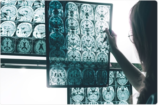
Blood Flow Through the Brain & Alzheimer’s Disease
Alzheimer’s disease is characterized by the presence of beta-amyloid plaques and neurofibrillary tangles of tau, that together lead to the progressive neurodegeneration of neurons in the brain leading to the symptoms of dementia.

Image Credit:Atthapon Raksthaput
Recent research has revealed that patients and animal models of Alzheimer’s disease display significant disturbances to brain blood flow from early on in disease-course. This article will highlight the importance of brain blood flow to health and how Alzheimer’s disease is marked with severe reductions.
Given that many traditional therapeutic approaches have failed in the past, perhaps brain blood flow-related therapies may pave the way to more successful treatments in the future.
Blood Flow to the Brain
The brain accounts for just 2% of total body weight but receives 20% of all the blood flow in the body from the heart every minute.
Brain cells (neurons) require vast amounts of energy to function properly and to maintain the metabolic demands neuronal activity exerts, the brain has an intricate system called neurovascular coupling.
Neurovascular coupling ensures that active brain regions receive a proportionally matched blood supply by increasing local cerebral blood flow (CBF).
Neurovascular coupling is facilitated by various cell types within the brain including neurons, astrocytes, pericytes, endothelial cells, smooth muscle cells, interneurons, and others.
Signaling between neurons and these cells eventually leads to increased CBF at both the capillary level as well as at the surface arterial level. Damage to specific cell types or signaling pathways could be the cause of neurovascular decline in aging and dementia.
Cerebral Blood Flow in Alzheimer’s Disease
Cerebrovascular dysfunction is implicated in the development and onset of dementia, including Alzheimer’s disease and vascular dementia.
The biggest risk factors that may compromise vascular function in Alzheimer’s disease include atherosclerosis (hardening and narrowing of arteries), hypertension (high blood pressure), diabetes and obesity.
These risk factors are the same for the development of cardiovascular diseases and cerebrovascular diseases including stroke. Many of these diseases have similar mechanisms, including oxidative stress and inflammation.
In addition to the vascular risk factors for the development of Alzheimer’s disease, it is known that CBF can be reduced as much as 25% if not more in patients with Alzheimer’s disease, known as chronic hypoperfusion (long term reduced blood flow).
The mechanisms behind this are still largely not fully understood, though impairments to neurovascular coupling as well as to the prolonged constriction of brain blood vessels and density could also be important.
Chronic hypoperfusion over time can lead to reduced oxygen delivery to brain tissue, causing neurons to become stressed and eventually die. This is in parallel to the neurodegenerative decline in neuronal numbers in the cortex as initiated by amyloid-beta plaques and tau tangles.
Furthermore, amyloid-beta deposits can form around blood vessels in the brain in what is known as cerebral amyloid angiopathy (CAA) that can also affect vessel reactivity (ability to vessels to dilate) and to cause vasoconstriction (constricted blood vessels).
Also, as the brain clears away amyloid-beta by clearance pathways that operate with functional blood flow, impairments to blood flow can lead to reduced clearance of amyloid-beta, causing it to accumulate in the brain, further exacerbating the condition.
Animal Studies of CBF in Alzheimer’s Disease – Potential Therapies for the Future?
Most of our knowledge and understanding of the mechanism underlying Alzheimer’s disease onset and progression have come from various animal studies using predominantly mouse models of the human condition.
For example, both humans and mice harboring the APOE4 allele (a known genetic risk factor for Alzheimer’s), show the breaking down of the blood-brain-barrier (BBB), which is attributed to the degeneration of a particular brain cell type called pericytes.
Alzheimer’s patients and mouse models have up to 50% reduction in pericytes that corresponds to the loss of approximately 350mm/mm3 capillary length.
A recent study from Chris Schaffer’s lab, published in Nature Neuroscience (2019), found a novel mechanism by which blood cells attaching to brain blood vessels leads to reduced CBF and impairs memory function in a mouse model.
A double transgenic mouse model harboring mutation to amyloid precursor protein (APP) and presenilin-1 (PSEN1) as well as another more severe model harboring 5 different mutations to APP & PSEN1 were studied.
The mutations within the transgenic mouse models are based on human familial mutations that cause Alzheimer’s disease. As such, these mouse models are used to better understand human Alzheimer’s disease with human genomic mutations.
Firstly, they assessed the function and structure of brain capillaries and found that the APP/PS1 double transgenic mouse model had a much higher fraction of capillaries with stalled blood flow, without any obstructions in the arterioles.
The stalled blood flow in these capillaries did not correlate with amyloid-beta/CAA, but when labeled for other body cell types, they found that areas of stalled blood flow were marked by the presence of leukocytes (white blood cells) attached to these blood vessels. At baseline, average CBF in these mice was 17% lower than healthy controls.
Researchers found that by injecting antibodies against these leukocytes (α-Ly6G) reduced the number of stalled capillaries within 10-15 minutes and increased CBF by around 13% in these mice by increasing flow rather than increasing vessel diameter (irrespective of any potential CAA).
This antibody led to the near-total depletion of leukocytes attached to brain capillaries. When subjected to cognitive memory tests, α-Ly6G injection led to improvements in object recognition tasks (working memory), but no changes to depression or motor function.
Finally, α-Ly6G injection also led to the reduced concentration of smaller amyloid-beta fragments in the brain by enhancing clearance pathways due to improved CBF, however, plaques themselves remained unchanged.
This study shows not only a novel mechanism of reduced blood flow in the brain due to Alzheimer’s disease by the attachment of leukocytes to capillaries but also that by blocking their attachment to vessels using antibodies, the reduced blood flow/stalling can be reduced to cause improvements in cognitive function.
The increased attachment of leukocytes to vessels may be due to the increased neuroinflammation leading to increased receptors on blood vessels. As such, this study highlights a novel therapeutic strategy.
In summary, cerebral blood flow (CBF) impairments, as well as impairments to the regulation of CBF in the brain (neurovascular coupling), are now becoming increasingly more evident as a mechanism in the development and progression of Alzheimer’s disease.
Recent research of both clinical and animal studies has revealed different mechanisms that could underpin this blood flow dysfunction in dementia, including a recent study revealing the adhesion of white blood cells to brain vessels to cause reduced CBF.
These studies have also revealed novel therapeutic targets for the potential treatment of dementia. Whilst they show promising results in mice, much more research in the form of clinical trials is needed in human patients before we can assume the success of such strategies.
Sources:
- Shabir et al, 2018. Neurovascular dysfunction in vascular dementia, Alzheimer’s and atherosclerosis. BMC Neurosci. 19: 62. pubmed.ncbi.nlm.nih.gov/…/
- Cruz Hernandez et al, 2019. Neutrophil adhesion in brain capillaries reduces cortical blood flow and impairs memory function in Alzheimer’s disease mouse models. Nature Neuroscience. 22:413–420. https://www.nature.com/articles/s41593-018-0329-4
Further Reading
- All Alzheimer's Disease Content
- Alzheimer’s Disease | Definition, Causes, Diagnosis & Treatment
- Alzheimer’s Disease Causes
- Alzheimer’s Disease Symptoms
- Alzheimer’s Disease Diagnosis
Last Updated: Mar 30, 2020

Written by
Osman Shabir
Osman is a Neuroscience PhD Research Student at the University of Sheffield studying the impact of cardiovascular disease and Alzheimer's disease on neurovascular coupling using pre-clinical models and neuroimaging techniques.
Source: Read Full Article


