What is Dual-Energy Computed Tomography?
Diagnostic imaging has been a widely used tool in the detection and treatment of various diseases. Dual-energy computed tomography (DECT) is an emerging imaging technique that combines information collected with various x-ray energy fields.
What is computed tomography?
Computed tomography (CT) is an imaging technique that utilizes a special x-ray apparatus to produce detailed images of the different parts of the body. Also dubbed as computerized tomography or computerized axial tomography (CAT), the technology has improved the detail and speed with which internal body structures can be evaluated. The entire series of photos made by CT scan can be viewed in 2-D and 3-D. However, most modern machines capture continuous photos in a spiral or helical way rather than getting a series of images of individual slices of the organs or body parts.
What is a dual-energy computed tomography (DECT)?
Dual-energy computed tomography (DECT) produces accurate anatomic and functional photos by manipulating the differences in the interactions of high- and low-energy photon spectra with various materials’ and tissues’ atomic factors to accurately discriminate the chemistry of tissues of the body as well as disease processes.
Standard computed tomography (CT) scanners utilize normal X-rays to produce cross-sectional images of body parts. Dual-energy CT uses both normal X-ray and a second one to produce more precise images.
What are the uses of dual-energy computed tomography (DECT) scan?
Diagnosis is an important means of detecting diseases and initiating treatment. Early diagnosis is the key to a successful treatment regimen. Recent developments and innovations in computed tomography have shown promise in data acquisition and in the analysis of data, to appropriately detect and diagnose diseases. With dual-energy computed tomography (DECT), it’s easier to get accurate images, showing abnormalities in various body parts.
It has become possible to obtain virtual unenhanced images from monochromatic reconstructions. Also, it has been widely used in detecting cardiovascular, hepatic, renal, adrenal, and pancreatic abnormalities.
Why do doctors recommend dual-energy computed tomography (DECT) scan?
Doctors can refer the patient for DECT scan because of many reasons. First, the DECT scanner can decrease or increase the effects of some chemical substances in the body. As a result, abnormalities can be seen more clearly. The images will show these abnormalities in more accurate photos. For instance, iodine, a common contrast media can show better images of blood vessels.
Furthermore, for body parts scanned without any contrast media or agents, the images can be produced in just a single examination, instead of the typical two separate examinations. The DECT can also improve image quality for patients with metal within the body (eg, joint replacement or debris from injury).
Lastly, the DECT can detect specific substances in the body that can help patients diagnosed with kidney stones to detect what type of stone is present. Hence, it can help in the appropriate treatment of the disease.
Patient information about dual-energy computed tomography (DECT)
What to expect?
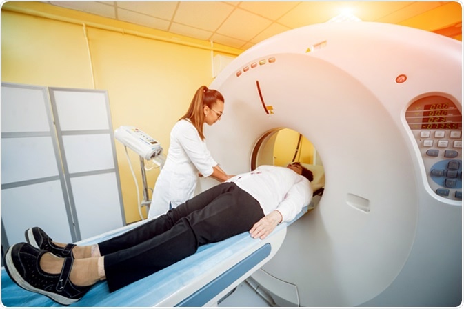
When you’re going to have a DECT, the patient will be asked to lie on a table. Depending on which part of the body is scanned, the patient may be asked to stay in various positions such as lying on the stomach, the back, or sides. Then, the table moves into the CT scanner, where an x-ray beam will rotate around the patient.
In some instances, the diagnostic procedure may require a special dye, dubbed as a contrast material or media, that works by highlighting an area to produce a clearer image. Usually, patients may receive the contrast media through a vein on the hands or they may be asked to drink it.
Preparation for the test
The prerequisites for DECT are almost the same with a standard CT scan. Pregnant patients should generally not have the scan unless the benefits of testing outweigh the risks to the developing fetus. For those who will need a contrast media, renal function should be assessed. This is important especially for patients who have diabetes, impaired kidney function, renal disease, those on the nephrotoxic drug, people over 60 years old, and those who are allergic to the substance.
Diagnostic scans have helped doctors to diagnose diseases. Early detection is essential to properly and promptly initiate the correct treatment. The prognosis for most diseases improves with early detection..
Sources
- https://www.ncbi.nlm.nih.gov/pubmed/25739115
- https://www.ncbi.nlm.nih.gov/pubmed/30219188
- https://www.sciencedirect.com/science/article/pii/S2212478017300254
- https://link.springer.com/article/10.3938/jkps.71.1056
- https://www.ncbi.nlm.nih.gov/pubmed/28711200
- https://www.ncbi.nlm.nih.gov/pubmed/28372824
- www.nibib.nih.gov/…/computed-tomography-ct
Further Reading
- All Tomography Content
- What is Optical Coherence Tomography?
- When is Optical Coherence Tomography Used?
- Micro CT Applications in Dentistry
- Whole-Body Low-Dose Computed Tomography (WBLDCT)
Last Updated: Apr 3, 2019

Written by
Angela Betsaida B. Laguipo
Angela is a nurse by profession and a writer by heart. She graduated with honors (Cum Laude) for her Bachelor of Nursing degree at the University of Baguio, Philippines. She is currently completing her Master's Degree where she specialized in Maternal and Child Nursing and worked as a clinical instructor and educator in the School of Nursing at the University of Baguio.
Source: Read Full Article


