Severe COVID-19 characterized by phosphorylation of STAT1
When a virus infects an individual, a complex human immune response is initiated to limit the effects of the viral invasion. One of the several different immune responses that can arise following a viral infection includes the generation of the pleiotropic cytokine interferon (IFN), which acts as a link between the innate and adaptive immune systems.
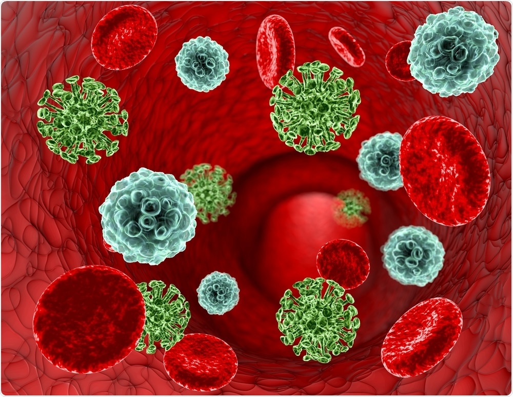
 Study: Altered increase in STAT1 expression and phosphorylation in severe COVID-19. Image Credit: Illustration Forest / Shutterstock.com
Antiviral effects of IFN
Previous studies have revealed that IFN-α, which is a type I IFN, is generated by plasmacytoid dendritic cells (pDCs). Comparatively, IFN-γ, which is a type II IFN, is secreted by natural killer (NK) cells, specific T-cells, and macrophages.
Both types of IFNs possess antiviral effects. Some examples of the activities elicited by these substances include the induction of apoptosis, as well as the activation of macrophages, NK cells, and both B- and T-lymphocytes.
Several studies have also been conducted to understand the IFN-related antiviral responses, particularly those that are involved in post severe acute respiratory syndrome coronavirus type 2 (SARS-CoV-2) infections. SARS-CoV-2 is a highly infectious ribonucleic acid (RNA) virus that is the causal agent of the coronavirus disease 2019 (COVID-19).
Inborn errors of type I IFN and the presence of autoantibodies against type I IFN appear to be associated with severe COVID-19 disease. In fact, previous reports indicate that approximately 10% of severely infected COVID-19 patients produce neutralizing autoantibodies against IFN-α, IFN-ω, or both.
Researchers have also found a highly impaired IFN type I signature, with decreased IFN-α production and activity, in severe COVID-19 patients. However, patients with mild cases of COVID-19 do not appear to experience these IFN-α production impairments or detectable levels of autoantibodies.
Phosphorylation in COVID-19
The Janus kinase (JAK)-signal transducer and activator of transcription (STAT) pathway has been instrumental in studies on gene expression and type I IFN signals. The JAK-STAT signaling pathway provides a highly complex and well-arranged system of heterogeneous molecules containing certain signaling receptor complexes, with the enlistment of different STATs that lead to specific downstream transcription. Additionally, the JAK-STAT pathway is a common signaling route of cytokines synthesis.
Scientists have found that a complex is formed after binding to the IFN-α receptor (IFNAR), which contains two chains including IFNAR1 and IFNAR2. This newly developed complex promotes phosphorylation of the receptor-associated JAK1 and tyrosine kinase 2 (TYK2).
Consequently, more phosphorylation of cytoplasmatic STAT1 and STAT2 leads to dimerization, interaction with IFN regulatory factor 9 (IRF9), as well as a strong enhancement of translocation to the nucleus. Tyrosine phosphorylation on Y701 occurs after the activation of STAT1, followed by nuclear accumulation.
Scientists have also shown that the viral proteins of NSP5, ORF7a, N, and ORF6 can directly interfere with JAK-STAT signaling either by blocking translocation of STATs or inhibiting their phosphorylation in SARS-CoV-2 infected cells. These studies have supported the hypothesis that an imbalance in JAK-STAT signaling is associated with COVID-19 severity.
About the study
A new study published on the preprint server medRxiv* reports an enhanced expression of STAT1 in mild and severely infected COVID-19 patients as compared to controls. Additionally, this study found a lowering in the expression of STAT1 in severely infected COVID-19 patients as compared to patients with mild infection. Interestingly, this altered expression was observed in plasmablasts and monocytes, both of which are chiefly associated with the pathogenesis of severe COVID-19 infection.
The reduced expression of STAT1 is complemented with an increase in the phosphorylation of STAT1 at pY701. These results indicate that disruption of JAK-STAT signal transduction is linked to reduced STAT1 transcription.
The researchers of the current study also found that an increased pSTAT1 occurred without stimulation in cultured CD19+ B-cells and CD3+ cells. This condition prevailed with additional IFN-α or IFN-γ stimulation.
The current study described upregulation of STAT1 and IRF9 in mildly and severely infected COVID-19 patients. This upregulation correlated with the IFN-signature reflected by Siglec-1 surface expression, which aligns with previous observations that Siglec-1 expression correlates with viral load in mild and severe COVID-19 patients.
This study also showed that the antiviral role of STAT1 is not entirely dependent on phosphorylation. In fact, the researchers observed the influence of alternative unphosphorylated STAT1 mediated pathways in COVID-19 infection.
The authors also routinely measured Siglec-1 in all COVID-19 patients who were admitted to intensive care units (ICUs) to differentiate patients with high and low IFN signatures. Like STAT1, Siglec-1 levels were reduced in severe COVID-19 patients as compared to those with mild cases.
Optimal treatment of COVID-19 patients
To date, the optimal treatment of patients with COVID-19 has not been determined. As a result, COVID-19 patients receive a wide range of treatments that often reduce IFN signaling by JAK-STAT inhibition. Comparatively, several types of IFN agents can also be used to treat COVID-19, whose efficacy has been tested in small clinical trials.
The current study revealed that patients with high IFN signaling are mostly in an early stage of the disease; therefore, inhibition strategies could be effective in these patients during the cytokine storm. For patients who experience a reduced antiviral response and are suffering from severe infection, IFN substitution may be a more promising approach as compared to further inhibition.
*Important notice
medRxiv publishes preliminary scientific reports that are not peer-reviewed and, therefore, should not be regarded as conclusive, guide clinical practice/health-related behavior, or treated as established information.
- Rincon-Arevalo, H., Aue, A., Ritter, J., et al (2021) Altered increase in STAT1 expression and phosphorylation in severe COVID-19. medRxiv. doi:10.1101/2021.08.13.21262006. https://www.medrxiv.org/content/10.1101/2021.08.13.21262006v1
Posted in: Medical Research News | Medical Condition News | Disease/Infection News
Tags: Apoptosis, Autoantibodies, CD3, Coronavirus, Coronavirus Disease COVID-19, Cytokine, Cytokines, Efficacy, Gene, Gene Expression, Immune Response, Intensive Care, Kinase, Phosphorylation, Receptor, Respiratory, Ribonucleic Acid, RNA, SARS, SARS-CoV-2, Severe Acute Respiratory, Severe Acute Respiratory Syndrome, Signaling Pathway, Syndrome, Transcription, Tyrosine, Virus

Written by
Dr. Priyom Bose
Priyom holds a Ph.D. in Plant Biology and Biotechnology from the University of Madras, India. She is an active researcher and an experienced science writer. Priyom has also co-authored several original research articles that have been published in reputed peer-reviewed journals. She is also an avid reader and an amateur photographer.
Source: Read Full Article


