Urine Analysis / Urinalysis
Urine analysis, also known as urinalysis, encompasses all the analytical tests carried out on a urine sample. It usually involves three forms of analysis:
- Physical – Assessment of the color, cloudiness and concentration of the urine
- Chemical – Examination of the chemical constituents of the urine using a test strip
- Microscopic – this allows one to look for bacteria, cells and cell fragments
Purpose
Urine analysis may be required as part of:
- Overall health assessment: for routine medical examination, pre-surgery preparation, or upon hospital admission to screen for disorders including kidney and liver disease.
- Medical Diagnosis: for those experiencing symptoms including abdominal pain, back pain, frequent or painful urination and blood in your urine.
- Medical Monitoring: following the diagnosis of kidney disease to track both condition and treatment.
- Specific testing. This includes pregnancy tests and drug screenings e.g. pregnancy tests measure the hormone human chorionic gonadotropin (HCG).
Urine analysis encompasses the following testing procedures:
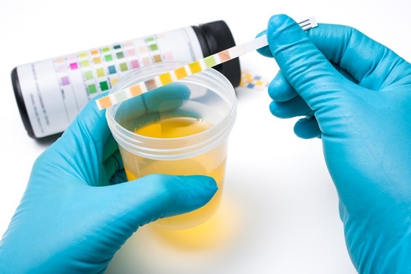
Rapid Urine Test
This test involves dipping a test strip embedded with colored fields into a urine sample for a few seconds. Depending on the concentration of the particular substance being testing for, the fields on the test strip change color. By using the color table on the test package as reference, one can see if any compounds exhibit abnormal urinary levels.
The following can be analysed:
- Colour: Fluid balance, diet, medicines, and diseases can affect this (e.g. Vitamin B, consumption of blackberries and beets).
- Clarity: Substances including bacteria, blood, or sperm can make normal, clear urine cloudy.
- Odor: Some diseases may cause a change in the odour of urine( e.g. diabetes can cause a sweet, fruity odour.)
- Specific gravity: Indicates production of urine by checking the concentration of substances in the urine against water content (e.g. when fluid intake is high, the kidneys produce urine with a high level of water, which has a low specific gravity.)
- pH: normally ranges from approximately 5 to 7 but certain treatments affect this and treatment may be needed to prevent the formation of some types of kidney stones.
- Protein: not normally present in the urine, but strenuous exercise, pregnancy, fever and some diseases (e.g. kidney disease and nephritis) may cause its appearance.
- Glucose: normally there is very little or no glucose in urine. When the kidneys are damaged and/or blood sugar level is very high (uncontrolled diabetes), urinary level increases.
- Nitrites: bacteria that cause a urinary tract infection (UTI) possess an enzyme that converts nitrates to nitrites. Thus, urinary nitrites indicate a UTI.
- Ketones: Fat is metabolised into ketones, which are expelled via urine. Large amounts of ketones in the urine may indicate diabetic ketoacidosis, a diet low in sugars and carbohydrates or starvation.
Microscopic Analysis
This involves centrifugation of a urine sample. Following this, the obtained sediment is spread on a slide and analysed microscopically to study components such as:
- Red or white blood cells: May be present in urine in disease, infection, or injury to the kidneys, ureters, bladder, or urethra.
- Casts: The formation of plugs of blood cells, proteins or other substances (casts) in the renal tubules that can occur in some renal diseases
- Crystals: Few crystals are found in healthy urine and many crystals, or certain types, may indicate presence of kidney stones or metabolic dysfunction.
- Bacteria, yeast cells, or parasites: Presence in urine indicative of an infection.
As with all tests, the results of urine tests are not always reliable. For this reason, a more detailed laboratory test is suggested following which abnormal results can be discussed with a doctor.
Urine Culture
This can be used to detect bacteria in the urine. A sample of midstream urine is incubated for 1-2 days with an appropriate medium to allow for bacterial growth. If any bacteria or fungi are present, there will be colony growth that will be studied based on size, form and color. Often urine cultures are used to test for a UTI then appropriate antibiotics are prescribed for its management.
References
- https://www.kidney.org/kidneydisease/twosimpletests
- http://www.ncbi.nlm.nih.gov/pubmedhealth/PMH0072534/
- http://library.med.utah.edu/WebPath/TUTORIAL/URINE/URINE.html
- http://www.healthlinkbc.ca/medicaltests/content.asp?hwid=hw6580
- http://patient.info/doctor/urine-dipstick-analysis
- http://www.highlands.edu/academics/divisions/scipe/biology/labs/cartersville/2122/urinalysis.htm
Further Reading
- All Stye Content
- Stye / Hordeolum – Painful Lumps on Eyelids
- What Causes a Stye?
- How to Treat a Stye
Last Updated: Feb 27, 2019

Written by
Afsaneh Khetrapal
Afsaneh graduated from Warwick University with a First class honours degree in Biomedical science. During her time here her love for neuroscience and scientific journalism only grew and have now steered her into a career with the journal, Scientific Reports under Springer Nature. Of course, she isn’t always immersed in all things science and literary; her free time involves a lot of oil painting and beach-side walks too.
Source: Read Full Article


