An immune flaw may cause West Nile viruss deadliest symptoms

Four out of five of people infected with the mosquito-borne West Nile virus (WNV) won’t even know it—heartening news when you consider there’s no vaccine to prevent the disease nor targeted medications to treat it. However, the rest can develop a serious illness—particularly the approximately 1% who get encephalitis, an inflammation of the brain that requires hospitalization. Up to 20% of these patients die.
What is it that makes these select few so vulnerable? An international team of researchers led by Rockefeller University’s Jean-Laurent Casanova and Alessandro Borghesi of the San Matteo Research Hospital in Pavia, Italy, has recently pinpointed the cause: an autoimmune defect that prevents the body from defending itself.
As published in the Journal of Experimental Medicine, they found that about 35% of patients hospitalized for WNV also carry autoantibodies that neutralize type 1 interferons, the signaling proteins deployed by a variety of cells in the fight against viruses. The prevalence was highest for people with encephalitis, 40% of whom had the autoantibodies that curb their own defense system from combating the viral invader.
West Nile virus now joins a small but important group of infectious diseases with a documented link between interferon-neutralizing autoantibodies and cases resulting in the most severe or deadly symptoms, including influenza (5%), COVID (15%), and MERS pneumonia (25%).
But with 40% of West Nile virus encephalitis cases now explained by this autoimmune deficiency, Casanova says, “West Nile virus is by far the best understood human infectious disease in the world. It’s stunning.”
From Uganda to everywhere
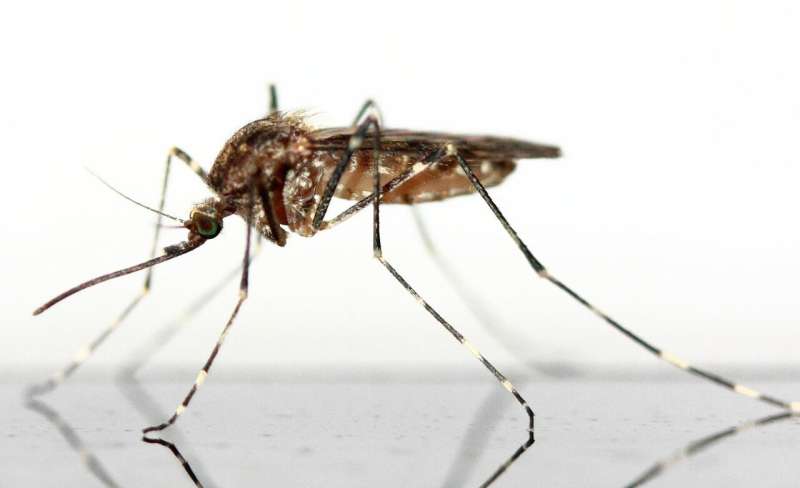
Since being discovered in the West Nile region of northwest Uganda in 1937, WNV has spread to 60 countries, largely following the migratory routes of birds. Culex mosquitoes, which are found virtually worldwide, pick up the virus from infected birds and pass it along to other animals, including us. Since the first cases in the U.S. were discovered in 1999 in New York, the disease has become the leading cause of mosquito-borne disease in the U.S. On the other side of the Atlantic, infection rates are similar across the northern Mediterranean, central Europe, and the Balkans.
Flaviviruses like WNV are single-stranded RNA viruses that are primarily found in ticks and mosquitoes, and infect people globally. They cause a range of serious diseases including dengue, Zika, and yellow fever.
In a small 2021 study, Casanova and his collaborators documented that three of eight people who had had adverse reactions to the yellow fever live-attenuated vaccine had autoantibodies to type 1 interferons as well. It made the team wonder whether this could be true for people severely affected by other injected viruses, also known as arboviruses; after all, “a mosquito’s proboscis is essentially the same idea as a syringe: It injects the virus directly into the bloodstream,” Casanova notes.
Moreover, a year before, he and his colleagues had found that about 15% of COVID cases were due to autoantibodies to type 1 interferons. They also knew that both the risk of encephalitis and potential to have autoantibodies increase with age. Perhaps these autoantibodies were responsible for severe outcomes in a variety of infectious diseases, no matter the method of delivery. “For all these reasons, we tested the hypothesis that perhaps some patients with West Nile encephalitis have these autoantibodies,” Casanova says.
Interfering with interferons
To find out, they studied blood, sera, plasma, or cerebrospinal fluid samples from 441 people who’d been hospitalized for life-threatening West Nile infections in three countries—the U.S., Italy, and Hungary—during viral outbreaks going back to 2002. Two-thirds were male, and the average age was 67. Most had been stricken with encephalitis, but some had meningitis (an inflammation of the lining of the brain or spine) or another neurological syndrome.
They tested samples for the presence of type 1 interferon-attacking autoantibodies, and then compared the results to people who had donated blood without knowing they were infected with WNV, which was detected during routine screening, and who had no or mild symptoms at a follow-up appointment.
Their results proved the team’s hunch: They discovered interferon-neutralizing autoantibodies in more than a third of the hospitalized patients, including about 40% of patients with encephalitis. The rate in the general population is 1%. This number is much higher than in the few other infectious diseases with an established link between this inborn error of immunity and severe outcomes. The only comparable rate was found in the small study they’d done on adverse reactions to yellow fever vaccine, where a third of the patients had the autoantibodies.
A select group
Type 1 interferons are supposed to help prevent viruses from crossing the blood-brain barrier. That they would be implicated in severe WNV fit with the high percentage of serious neuroinvasive conditions manifesting in these patients.
But the researchers were able to pinpoint the problem even more specifically. Of the 16 subtypes of type 1 interferons, only two were affected: 12 IFN-α (alpha) and the single IFN-ω (omega). Casanova’s group suspects this could be due to the fact that alpha and omega types are—just as injected viruses are—making blood the viral battlefield.
That researchers detected these autoantibodies during the first few days of hospitalization, before the patients’ immune systems had time to produce antibodies against WNV, made clear that the virus’s real danger to these patients lies in its ability to opportunistically capitalize on a hidden glitch already lurking within their immune systems. “This is really evidence that the autoantibodies to interferon don’t result from West Nile virus infection but instead are a cause of viral encephalitis,” Casanova says.
Yet how the autoantibodies curb the interferons’ activity remains unclear, as does where that hindrance is taking place. “We don’t know whether the autoantibodies cause West Nile encephalitis by preventing the control of the virus in the blood or in the brain,” he says. “And we don’t know which cells are their targets.”
Prevention, testing, treatment
The findings have a variety of clinical implications. One of the researchers’ recommendations is to screen the general population for autoantibodies in areas where WNV is endemic, beginning with the elderly and people with autoimmune diseases. More broadly, such screenings could be useful for doctors treating patients with viral conditions, especially those caused by arboviruses such as dengue, yellow fever, tick-borne encephalitis, and Chikungunya.
They could also clue patients and clinicians into what they might be up against, should an infection take place. “If you know you have the autoantibodies and you develop symptoms, you can tell people in the emergency room about it, and that’s going to help the team manage you,” Casanova says.
Such management could potentially involve interferon treatments, which could be useful even for infected patients who don’t have autoantibodies. They’re currently used to treat conditions including chronic hepatitis C, some leukemias, and multiple sclerosis.
Other treatments would take years more research, Casanova says. “One idea is to think of ways to deplete the B cells and plasma cells that make these autoantibodies. Another possibility would be to perhaps use decoys—molecules that would lure the autoantibodies,” he says. “Those are very hypothetical, but there are possible therapeutic interventions on the horizon. Not now, and not next year—but on the horizon.”
More information:
Adrian Gervais et al, Autoantibodies neutralizing type I IFNs underlie West Nile virus encephalitis in ∼40% of patients, Journal of Experimental Medicine (2023). DOI: 10.1084/jem.20230661
Journal information:
Journal of Experimental Medicine
Source: Read Full Article

