What is iDISCO?
Visualizing Structures within Tissue
To see structure within biological tissue such as organs, it is necessary to first label the tissue. Antibodies are frequently used for this purpose, and this is known as “immunolabelling”.
Antibodies are specific to certain “markers”, or antigens, and immunolabelling is used for diagnosis and research. Changes in structure between treated, untreated but healthy, and diseased tissue can be seen with immunolabelling.
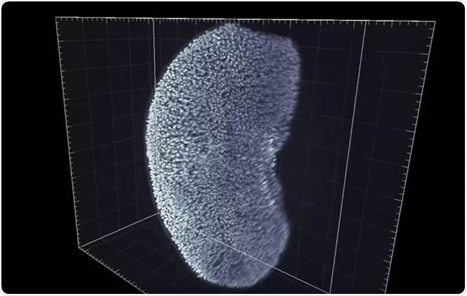
It is difficult to label deep within an intact tissue, meaning that a drawback of immunolabelling is that a thin layer of tissue is needed. 3D imaging is desirable to observe complex structures within tissues that cannot be imaged in 2D, including structures such as axonal tracts.
Immunolabelling-Enabled 3DISCO, or iDISCO
Immunolabelling Whole Tissues
Renier and co., the developers of iDISCO, initially looked at many processes that had already been developed to immunolabel whole tissues. They then combined some approaches to optimize the process. This included using non-ionic detergents, as well as dehydration-rehydration cycles using organic solvents, which remove membrane fats (lipids), and using hydrogen peroxide or methanol to stop the tissue sample from becoming fluorescent without any labels (autofluorescence). While methanol was good at eliminating autofluorescence, it also prevented some of the antibodies they tested from binding.
To reduce the background noise generated by immunolabelling, Renier and co. found that treating the tissue with glycine and heparin worked well. In addition, to counteract autofluorescence, using fluorescent dyes in the red end of the spectrum worked best due to the fact that autofluorescence is usually in the blue to green end of the spectrum.
Making Tissues Transparent
Renier and co. also established which of the methods developed to make biological tissue transparent best supported the immunolabelling procedure. The process needed to be able to clear tissues with good efficiency, be inexpensive and work with a wide variety of antigens. This included 3DISCO (3D Imaging of Solvent-cleared Organs), which was developed by Ertürk and co. Originally, 3DISCO was an improved version of the benzyl alcohol-benzyl benzoate (BABB) method, which cleared lipid-rich tissue such as nerve tissue.
The Process
Fixing the Samples
The tissue samples are initially fixed with 4% PFA in phosphate buffered saline (PBS). This is left overnight at 4°C, then another fixation step is carried out at room temperature for 1 hour using the same fixing agent.
Pre-Treatment
First, the fixed samples are washed twice with PBS for 1 hour per wash. To dehydrate the samples a 20% methanol solution is added, with the concentration being progressively increased to 40%, 60%, 80%, then 100%. The sample is left in each methanol solution for 1h. The sample is then cooled at 4°C. The tissue is then placed into a 66% dichloromethane (DCM):33% methanol solution overnight at room temperature. The sample is then washed twice more in 100% methanol, and cooled at 4°C. 5% hydrogen peroxide (H2O2) in methanol is then used to bleach the sample overnight at 4°C. To rehydrate the sample, the sample is placed into an 80% methanol solution, then 60%, 40% and 20% for 1h at each concentration. The sample is then washed twice using a PBS TritonX-100 solution for 1h per wash.
Alternatively, different chemicals can be used to pre-treat samples if methanol is not compatible, or if embryonic tissue or adult tissue which is less than 1 mm in thickness is to be used.
Immunolabelling
The pre-treated sample is then placed into the “permeabilization solution”, made up of PBS, TritonX-100, glycine and DMSO. This is incubated at 37°C for a maximum of 2 days, depending on the type of tissue. Then the sample is placed into a “blocking solution” comprising of PBS, TritonX-100, serum (which contains proteins) and DMSO. At this stage, proteins are used to prevent the antibodies from binding non-specifically. The tissue is incubated in this blocking solution at 37°C for up to 2 days, again depending on the type of tissue.
Antibodies are then added to the sample one at a time, in a solution of PBS, Tween-20, heparin serum and DMSO for the first antibody to be added. The tissue is then incubated at 37°C for a varying time, depending on the sample type. Before the addition of a second antibody, the sample is washed 4-5 times with PBS, Tween-20, and heparin.
Clearing
The sample is once again dehydrated using progressively more concentrated methanol solutions, ending with two periods of incubation with 100% methanol. The sample is then incubated for 3h with 66% DCM/33% methanol, then placed into 100% DCM twice for 15 minutes each. The sample is then finally incubated with dibenzyl ether before being imaged.
Sources
- Original iDISCO paper – www.cell.com/cell/fulltext/S0092-8674(14)01297-5#secsectitle0115
- iDISCO protocol – idiscodotinfo.files.wordpress.com/…/…rotocol-methanol-dec-2016.pdf
Further Reading
- All iDISCO Content
- iDISCO Applications
Last Updated: Sep 4, 2018

Written by
Dr. Maho Yokoyama
Dr. Maho Yokoyama is a researcher and science writer. She was awarded her Ph.D. from the University of Bath, UK, following a thesis in the field of Microbiology, where she applied functional genomics toStaphylococcus aureus . During her doctoral studies, Maho collaborated with other academics on several papers and even published some of her own work in peer-reviewed scientific journals. She also presented her work at academic conferences around the world.
Source: Read Full Article